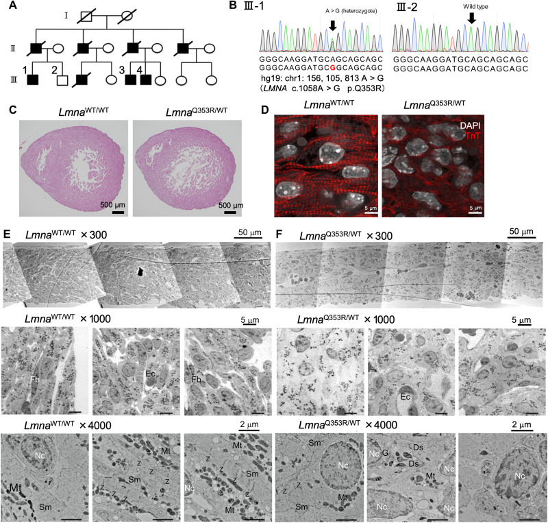
Fig. 1. Immature intracellular structure of LmnaQ353R/WT mice.
(A) Family tree of the DCM cohort with the LMNA mutation (p.Q353R). Shapes filled with black color indicate the patients with DCM. The squares indicate the men and the circles the women. Diagonal lines indicate the family members that have died. (B) Sanger sequencing of genomic DNA of a patient (III-1) and a healthy sibling in the cohort (III-2), including the mutation site. (C) Hematoxylin and eosin staining analysis of LmnaWT/WT and LmnaQ353R/WT knock-in murine hearts on embryonic day 17.5 (E17.5). (D) Immunostaining of troponin T (TnT) in LmnaWT/WT and LmnaQ353R/WT knock-in mice on E17.5. DAPI, 4′,6-diamidino-2-phenylindole. (E) Electron microscopic images showing structure of left ventricular wall in an LmnaWT/WT mouse on E17.5. Fb, fibroblast; Ec, endothelial cell; Nc, nucleus; Sm, sarcomere; Mt, mitochondria; Z, z-disc. (F) Electron microscopy images showing structure of left ventricular wall in an LmnaQ353R/WT knock-in mouse on E17.5. Ds, desmosome; G, Golgi body; WT, wild type.

