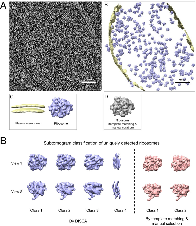
Fig. 4.
(A) Example unsupervised annotation on a Mycoplasma pneumoniae cell tomogram (65): a slice of the original tomogram; b detected patterns reembedded to the original tomogram space; c isosurface visualization of detected patterns identified (generated from subtomogram averaging); d isosurface visualization of the ribosome structure using the template-matching approach. (B) Relion subtomogram classification of uniquely detected ribosomes by the two approaches.

