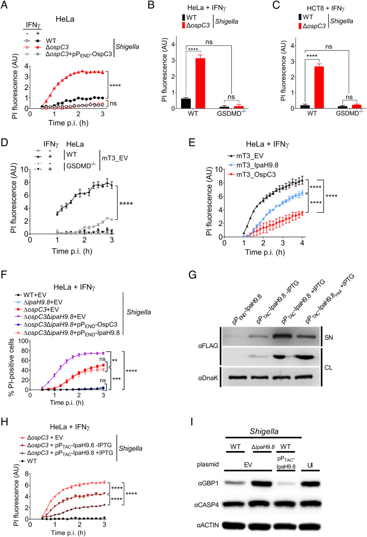
Fig. 1.
Shigella OspC3 and IpaH9.8 cooperate to suppress bacteria-triggered pyroptosis of IFNγ-primed epithelial cells. (A–F, H, and I) WT or GSDMD−/− HeLa or HCT8 cells unprimed or primed overnight with 10 ng/mL IFNγ were infected with the indicated strains at an MOI of 100 (A), 10 (B–F, and H), or 5 (I). Cells were infected with Shigella strains that carry pBAD33-AfaI and designated plasmids (B and C, H–I). For Shigella, 30 min postinfection (p.i.) (A–C, F, H, and I) and for mT3, 1 h p.i. (D and E), PI (A–E, and H) or PI and Hoechst (F) were added to the medium, and cell death was measured by monitoring PI upt,ake using a plate reader (A–E, and H) or PI and Hoechst uptake using an automated fluorescence microscope (F). A time course of cell death is shown in (A, and D–F) and a 3-h end point in (B). (G) Immunoblots of secretion assays of IpaH9.8-FLAG expressed from designated plasmids by ΔospC3 Shigella. SN (supernatant) and CL (cell lysate) fractions were probed with anti-FLAG to detect secreted and expressed effectors and DnaK, a cytosolic Shigella protein, as a lysis and loading control. (I) Immunoblots of lysates of IFNγ-primed HeLa cells infected with indicated Shigella strains probed with indicated antibodies. EV = empty vector, UI = uninfected, and AU = arbitrary units. Values shown are the mean ± SEM of three experimental replicates. Data were analyzed using two-way ANOVA with Tukey’s post hoc test. **P < 0.01, ***P < 0.001, ****P < 0.0001, ns = nonsignificant.

