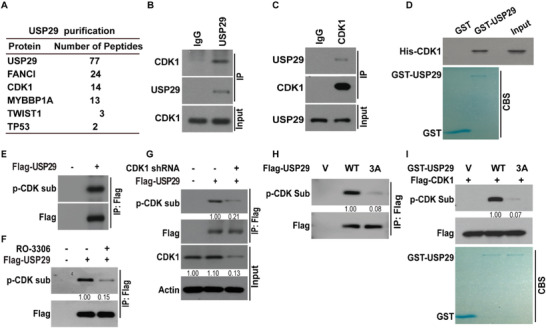
Figure 4.

CDK1 binds and phosphorylates USP29. A) List of USP29‐associated proteins identified by mass spectrometric analysis. MDA‐MB‐231 cells stably expressing Flag‐USP29 were generated and USP29 complexes were subjected to mass spectrometric analysis. MDA‐MB‐231 cell lysates were subjected to immunoprecipitation with B) IgG, anti‐USP29 or C) anti‐CDK1 antibody. The immunoprecipitates were blotted with indicated antibodies. D) Purified recombinant GST, GST‐USP29 and His‐CDK1 were incubated in vitro as indicated. The interaction between USP29 and CDK1 was examined. CBS, Coomassie blue staining. E) Empty vector or Flag‐USP29 were transfected in cells stably expressing USP29 shRNA. Cell lysates were subjected to immunoprecipitation with anti‐Flag antibody and the phosphorylation of USP29 was examined by phospho‐CDK Substrate (p‐CDK sub) antibody. F) Cells were transfected with indicated plasmids and treated with vehicle, or CDK1 inhibitor (RO‐3306). Cell lysates were subjected to immunoprecipitation with anti‐Flag antibody and the phosphorylation of USP29 was examined by phospho‐CDK Substrate antibody. G) Cells were transfected with indicated plasmids. Cell lysates were subjected to immunoprecipitation with anti‐Flag antibody and the phosphorylation of USP29 was examined by phospho‐CDK Substrate antibody. H) Empty vector or Flag‐USP29 (WT or 3A mutant) was transfected in MDA‐MB‐231 cells stably expressing USP29 shRNA. Cell lysates were subjected to immunoprecipitation with anti‐Flag antibody and the phosphorylation of USP29 was examined by phospho‐CDK Substrate antibody. I) CDK1 phosphorylates USP29 in vitro. Cell lysates of MDA‐MB‐231 cells stably expressing Flag‐CDK1 were immunoprecipitated with anti‐Flag antibody and incubated with bacterial purified GST, GST‐USP29 WT, or GST‐USP29 3A mutant. Western blot was performed and the phosphorylation of USP29 was examined by phospho‐CDK Substrate antibody.
