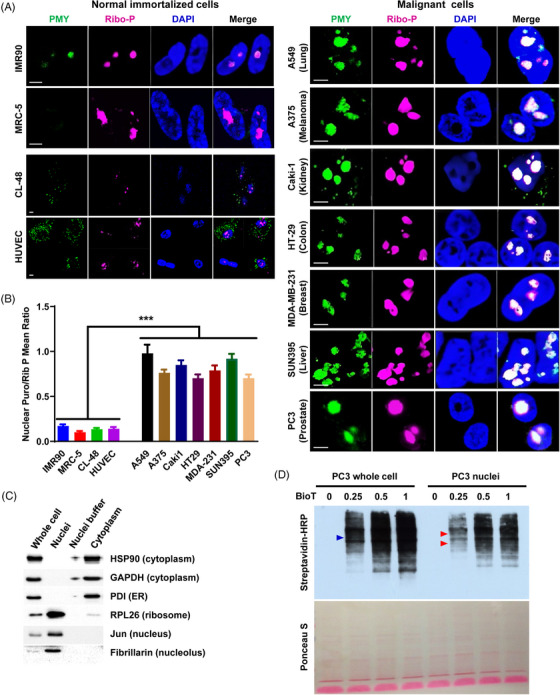
FIGURE 1.

Enhanced nuclear translation in malignant cancer cells. (A, B) NRPM analysis on normal immortalized cells (left panel) and malignant cancer cells (right panel) across seven cancer types as indicated (A). Scale bar: 5 µm. Twenty fields were acquired for each condition, and the mean fluorescence ratio of PMY/Ribosome P staining for each field was quantitated using ImageJ (B). (C) The purity of isolated PC3 nuclei was determined by western blotting using the antibodies against specific marker proteins as indicated. (D) Newly synthesized proteins from whole cells or isolated nuclei of PC3 cells were labeled by biotinylated Puromycin (BioT) and detected by western blotting using streptavidin‐horseradish peroxidase (HRP). Triangles indicate potential distinct proteins. Ponceau S staining is shown as a control. Data are shown as mean ± SEM. ***p < 0.005. Two‐tailed unpaired t‐test. PMY: puromycin; Ribo‐P: ribosomal P.
