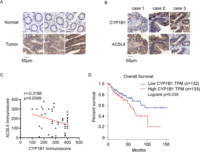
Fig. 5. The correlation between CYP1B1 and ACSL4.
A IHC staining of CYP1B1 in CRC cancer tissues and adjacent normal tissues. B IHC staining of CYP1B1 and ACSL4 in CRC cancer tissues. C The correlation analysis between CYP1B1 and ACSL4 in CRC cancer tissues was explored using Pearson correlation analysis (n = 44). D Overall survival was analyzed in COAD patients from the GEPIA database with low or high expression of CYP1B1 (defined by RNA sequencing with group cut-off values of 50 and 50%).

