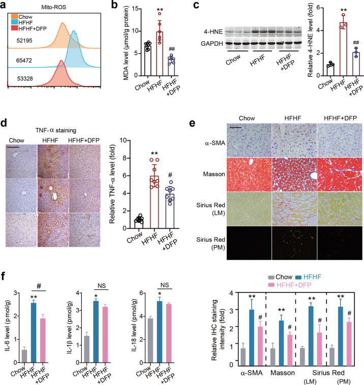
Fig. 8. An iron chelator DFP alleviates steatohepatitis.
a Mitochondrial ROS level was determined by flow cytometry with MitoSOX dye. b The MDA levels in liver tissues in three groups of mice. c Representative immunoblot images and quantitative analysis of 4-HNE protein level in liver tissues in three groups of mice. GAPDH as loading control. n = 3 biologically independent experiments. d Immunohistochemistry staining of TNF-α in liver tissues in three groups of mice. Scale bars, 100 μm. n = 8 biologically independent experiments. e Immunohistochemistry staining of α-SMA, Masson’s trichrome staining and Sirius Red staining (red under light microscopy [LM]; orange/green under polarized microscopy [PM]) in liver tissues in three groups of mice. Scale bars, 100 μm. f ELISA of IL-6, IL-1β and IL-18 levels in liver tissues of three groups of mice. The data were presented as Means ± SEM and analyzed by One way-ANOVA followed by Tukey’s post hoc. *P < 0.05, **P < 0.01 vs Chow; #P < 0.05, ##P < 0.01 vs HFHF. NS no significance.

