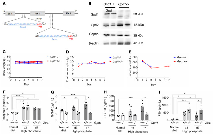
Figure 3. Gpd1 mediates phosphate-stimulated G-3-P and FGF23 production.
(A) Schema for targeted deletion of Gpd1. (B) Immunoblot of Gpd1, Gpd2, Gapdh, and β-actin in kidney tissue from Gpd1+/+ and Gpd1–/– mice. (C–E) Body weight (C), food consumption (D), and urine Pi (E) from Gpd1+/+ and Gpd1–/– mice on high phosphate diet (1.2%) for 7 days (note, food consumption and urine Pi were assessed per cage, n = 4 mice per diet group). (F–I) Blood phosphate (F), G-3-P (G), intact FGF23 (iFGF23) (H), and PTH (I) concentrations in Gpd1+/+ and Gpd1–/– mice fed a normal diet (0.6% Pi) and after 3 and 7 days on high phosphate diet (1.2% Pi) (n = 3–6 per group). Values are mean ± SEM. *P < 0.05, ***P < 0.0001. Unpaired student’s t test (F–I).

