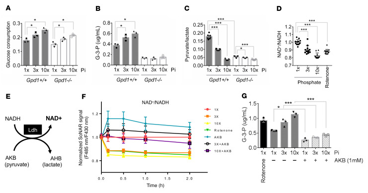
Figure 5. Phosphate-stimulated glycolysis and Gpd1 activity are coupled through cytosolic NAD+ recycling.
(A–C) Media glucose consumption (A), media G-3-P concentration (B), and media pyruvate/lactate ratio (C) from primary proximal tubular cells isolated from Gpd1+/+ and Gpd1–/– mice treated for 6 hours with increasing concentrations of phosphate (Pi) (n = 3 per group, with 3 technical replicates per sample for C). (D) Cytosolic NAD+/NADH measured in SoNar-expressing OK cells treated with rotenone (0.5 μM) or increasing concentrations of phosphate for 2 hours (n = 12 per group). (E) Schema for LDH-mediated NAD+ recycling; AKB, α-ketobutyrate; AHB, α-hydroxybutyrate. (F) Cytosolic NAD+/NADH measured with SoNar in OK cells treated with rotenone or increasing concentrations of phosphate ± AKB (1 mM) (n = 3 per group). (G) Media G-3-P concentrations from OK cells treated with rotenone (0.5 μM) or increasing concentrations of phosphate ± AKB (1 mM) for 2 hours (n = 3 per group). 1 × Pi = 0.9mM. Values are mean ± SEM. *P < 0.05, ***P < 0.0001. ANOVA with Dunnett’s multiple comparisons test (A–D) or ANOVA with Tukey’s multiple comparisons test (G).

