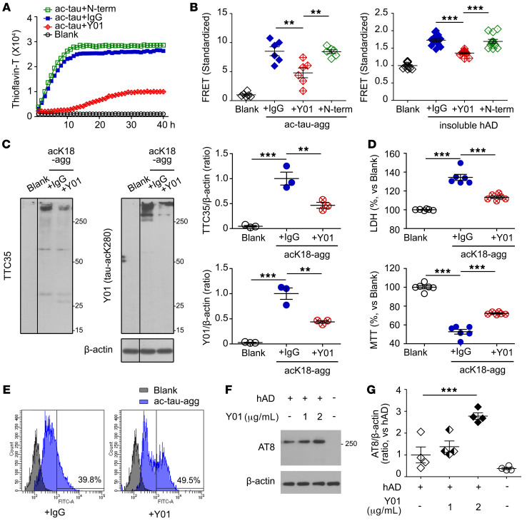
Figure 3. Y01 antibody inhibits acetylation-induced tau aggregation and propagation but enhances microglial tau uptake by Y01.
(A) Y01 antibody decreases aggregation of acetylated tau. IgG was used as a control. Anti–tau-N-term antibody does not decrease aggregation of acetylated tau. (B) Quantification of Y01-mediated inhibition of the FRET signal from HEK293T tau biosensor cells. Cells were pretreated with either Y01, anti–tau-N-term, or IgG, and tau seeding was induced by addition of either ac-tau-agg (n = 6 per group) or sarkosyl-insoluble fractions from human AD brain (n = 12 per group). (C) Y01 antibody decreased tau aggregation in primary mouse cortical neurons treated with acK18-agg. IgG was used as a control. The lanes were run on the same gel but were noncontiguous. AcK18-agg was analyzed by semidenatured immunoblotting using TTC35 or Y01 antibody, and aggregation was quantified by densitometry. n = 3 per condition. (D) Representative LDH and MTT assays of primary neurons treated with acK18-agg and control IgG or Y01. n = 6 per group. (E) Flow cytometric quantification of mean fluorescence intensity (arbitrary units) of primary mouse microglia treated with ac-tau-agg in the presence of control IgG or Y01. (F and G) BV2 microglial cells were treated with sarkosyl-soluble fractions from AD brains with either control IgG (2 μg/mL) or Y01 (1 or 2 μg/mL) for 4 hours. (F) Semidenaturing immunoblots with AT8 antibody. (G) Quantification of AT8 levels normalized to β-actin. n = 4 per group. Statistical analysis was performed by 1-way ANOVA followed by Tukey’s multiple-comparison test. **P < 0.01, ***P < 0.001. The error bars represent the SEM.

