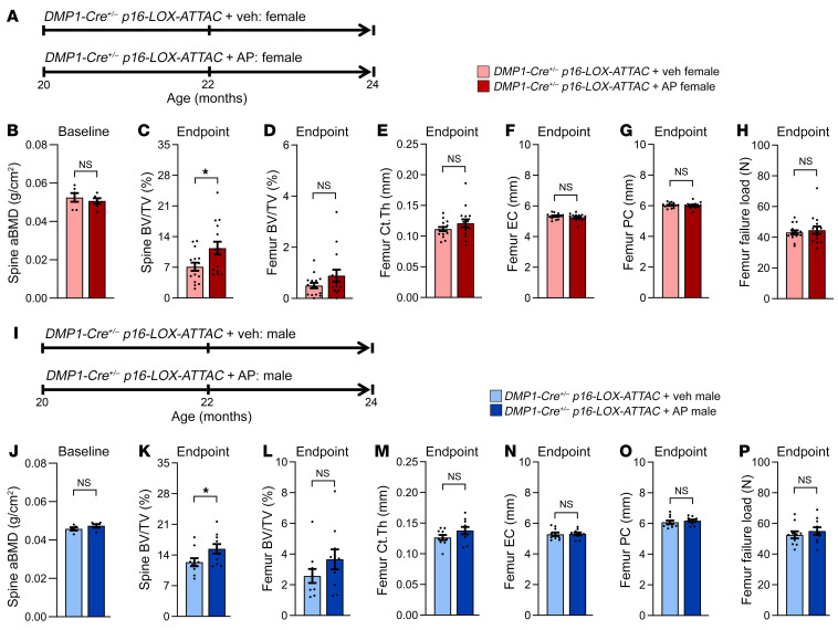
Figure 2. Effects of local Sn osteocyte–specific clearance on the skeleton of old female and male mice.
(A) Study design for local clearance of Sn osteocytes in female old (20 months) DMP1-Cre+/– p16-LOX-ATTAC mice randomized to Veh (pink) or AP (red) treatment for 4 months. (B) DXA-derived aBMD (g/cm2) at baseline in 20-month-old females (n = 6 females/group). (C) Quantification of the study endpoint (24 months) μCT-derived bone volume BV/TV fraction at the lumbar spine in mice treated with Veh (n = 15 females) versus AP (n = 15 females). (D–H) Quantification of μCT-derived (D) BV/TV, (E) cortical thickness (Ct.Th), (F) endocortical circumference (EC), (G) periosteal circumference (PC), and (H) μFEA-derived failure load at the femur metaphysis in female mice (n = 15 females/group). (I) Study design for local clearance of Sn osteocytes in old (20 months) male DMP1-Cre+/– p16-LOX-ATTAC mice randomized to Veh (light blue) or AP (dark blue) treatment for 4 months. (J) DXA-derived aBMD (g/cm2) at baseline in 20-month-old male mice (n = 5/males group). (K) Quantification of the study endpoint (24 months) μCT-derived BV/TV at the lumbar spine in male mice treated with Veh (n = 10 males) or AP (n = 10 males). (L–P) Quantification of μCT-derived (L) BV/TV, (M) cortical thickness, (N) endocortical circumference, (O) periosteal circumference, and (P) μFEA-derived failure load at the femur metaphysis in male mice (n = 10/group). Data represent the mean ± SEM. NS, P > 0.05; *P < 0.05 and **P < 0.01, by independent samples Student’s t test or Wilcoxon rank-sum test, as appropriate.

