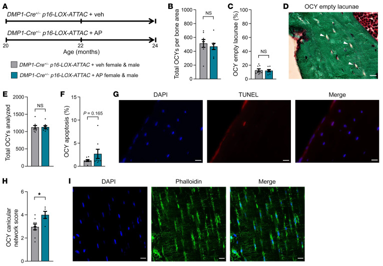
Figure 4. Effects of local Sn osteocyte clearance on empty lacunae, osteocyte apoptosis, and osteocyte LCN quality.
(A) Study design for the local clearance of Sn osteocytes in old (20 months) DMP1-Cre+/– p16-LOX-ATTAC mouse cohorts, males and females combined, randomized to Veh (gray) or AP (teal) treatment for 4 months. (B–D) Study endpoint (24 months) quantification of (B) total osteocytes per bone area and (C) percentage of osteocyte empty lacunae in mice treated with Veh (n = 8: n = 4 females, n = 4 males) versus AP (n = 8: n = 4 females, n = 4 males), and (D) representative images of osteocyte empty lacunae (arrowheads). Scale bar: 25 μm. (E–G) Quantification of osteocyte apoptosis, including (E) total numbers of osteocytes analyzed and (F) percentage of osteocyte apoptosis in mice treated with Veh (n = 8: n = 4 females, n = 4 males) versus AP (n = 8: n = 4 females, n = 4 males), and (G) representative images of DAPI-stained, TUNEL+, and merged apoptotic osteocytes. Scale bars: 25 μm. (H) Quantification of osteocyte LCN score for mice treated with Veh (n = 9: n = 5 females, n = 4 males) versus AP (n = 5: n = 3 females, n = 2 males), and (I) representative images of DAPI-stained, phalloidin-stained, and merged osteocyte LCNs. Scale bars: 25 μm. Data represent the mean ± SEM. NS, P > 0.05; *P < 0.05, by independent samples Student’s t test or Wilcoxon rank-sum test, as appropriate.

