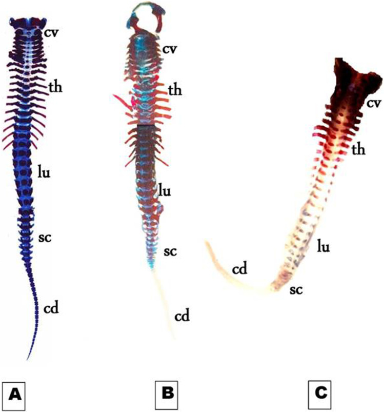
Fig. 5.
Photomicrographs of a ventral view of the vertebral column of the 19th day of gestation rat fetuses. A Control vertebral column showing well-ossified centra and parts of the neural arches of the cervical (cv), thoracic (th), lumbar (lu), sacral (sc), and the first third of caudal vertebrae (cd). B Vertebral column of fetus maternally treated with a low dose of MSG showing the absence of chondrification and ossification of the most caudal vertebra (cd). C Vertebral column of the fetus maternally treated with a high dose of MSG showing the absence of chondrification in all vertebrae and delay in ossification of the centra of the cervical vertebrae. Note that there is no ossification and chondrification of most caudal vertebrae (6.3X)

