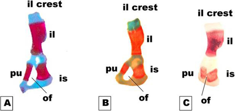
Fig. 8.
Photomicrographs of a dorsal view of the half of the pelvic girdle on the 19th day of gestation rat fetuses. A Control half pelvic girdle showing, well-developed ilium (i), ischium (is), and pubis (pu). B The half pelvic girdle of the fetus maternally treated with a low dose of MSG showed a normal appearance but with faint staining of both bone and cartilage. C The half pelvic girdle of the fetus maternally treated with a high dose of MSG showing a complete absence of chondrification and altered size of the ileum (il), ischium (is), and pubis (pu) (appear thicker and shorter than normal) and delayed obturator foramen (of) (10X)

