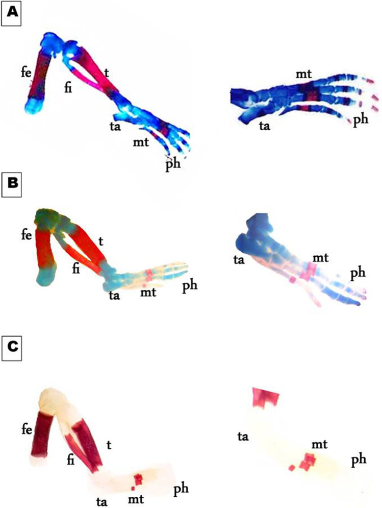
Fig. 9.
Photomicrographs of a lateral view of the hind limb of the 19th day of gestation rat fetuses. A Control hind limb showing, fully developed cartilage and bone of femur (fe), tibia (t), tarsals (tr), metatarsals (mt), and phalanges (ph). B Hind limb of the fetus maternally treated with a low dose of MSG shows a delay in chondrification of tarsals (ta) and phalanges (ph). C Hind limb of the fetus maternally treated with a high dose of MSG shows the complete absence of chondrification in the entire limb besides the absence of ossified phalanges (ph) (10X, 16 X)

