
Fig. 9.
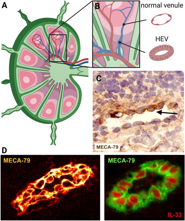
High endothelial venues (HEVs). A HEVs in secondary lymphoid organs present in the T-cell zone of the lymph node is the location with active extravasation of leukocytes. B HEVs display a cuboidal EC morphology. C An HEV in the inflamed synovium of a rheumatoid arthritis patient. D HEVs in human tonsils, stained for MECA-79 and the HEV nuclear cytokine IL-33 (right). Photomicrographs by courtesy of Drs. Blanchard and Girard, Toulouse, France [338]. Figure is created with BioRender.com and is available on request