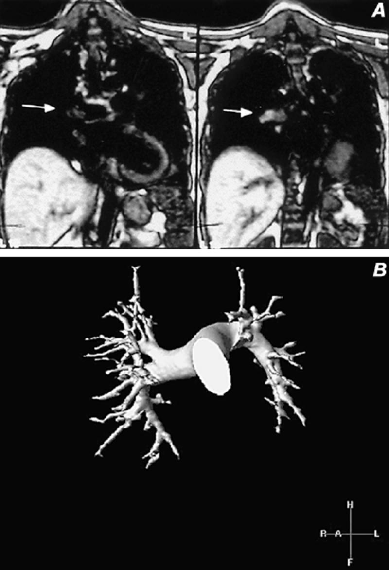
Fig. 10 Pulmonary artery magnetic resonance images. A) Coronal spin-echo image demonstrates an area of increased signal in an embolus within the right pulmonary artery extending into the intralobar artery (arrow) B) Volume-rendered contrast-enhanced study in a normal pulmonary arterial system. This 3-D technique enables rotation about all 3 axes in space to provide comprehensive evaluation of the entire pulmonary arterial system.
