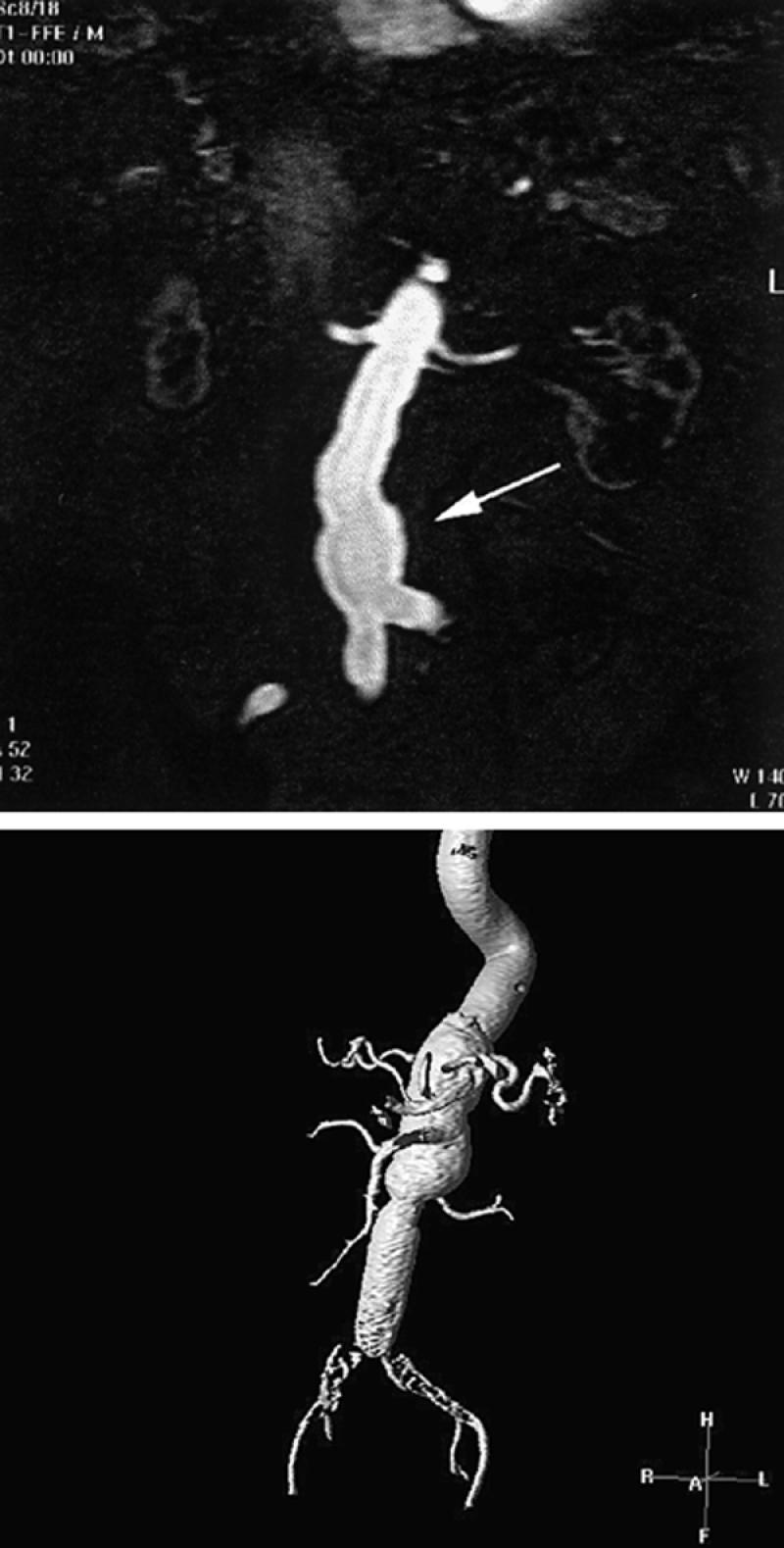
Fig. 12 Contrast-enhanced magnetic resonance angiogram of abdominal aortic aneurysms. A) Single 2-mm-thick coronal image discloses the proximal portions of the single renal arteries as well as opacification of a portion of the distal abdominal aortic aneurysm. Thirty to 40 of these contiguous images can be stacked together and displayed in a variety of 3-D views. These single images (source images) provide useful information on the status of branch vessels. B) Contrast-enhanced 3-D display (using volume rendering) of an abdominal aortic aneurysm in another patient. Typically, these images are rotated around the x, y, and z axes every 10 degrees, for comprehensive display of branch vessels. Note that large areas can be examined.
