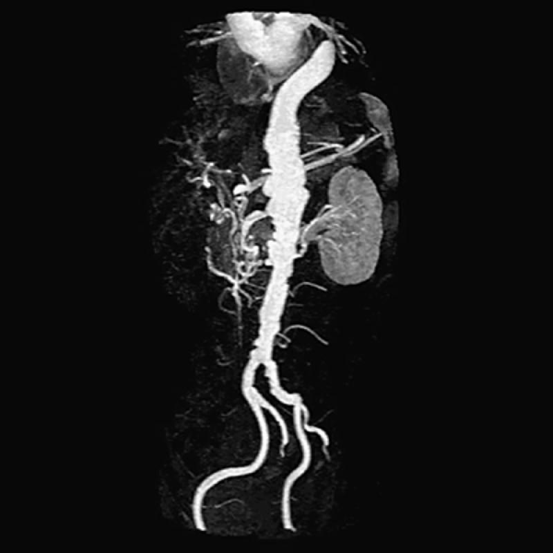
Fig. 15 Three-dimensional contrast-enhanced oblique image (maximum intensity projection) of 1st of 3 separate image set acquisitions (1 of the abdominal aorta and subsequent images obtained in the thighs and calves). Note poor delineation of the renal arteries and an ectasia of the aorta above the level of the kidneys. This is an oblique 3-D projection, which displays the tortuous but patent iliac vessels quite well.
Note: Contrast-enhanced images display only the patent lumen. In patients with abdominal aortic aneurysm, spin-echo images must also be obtained to delineate the outer limits of the aneurysmal wall.
