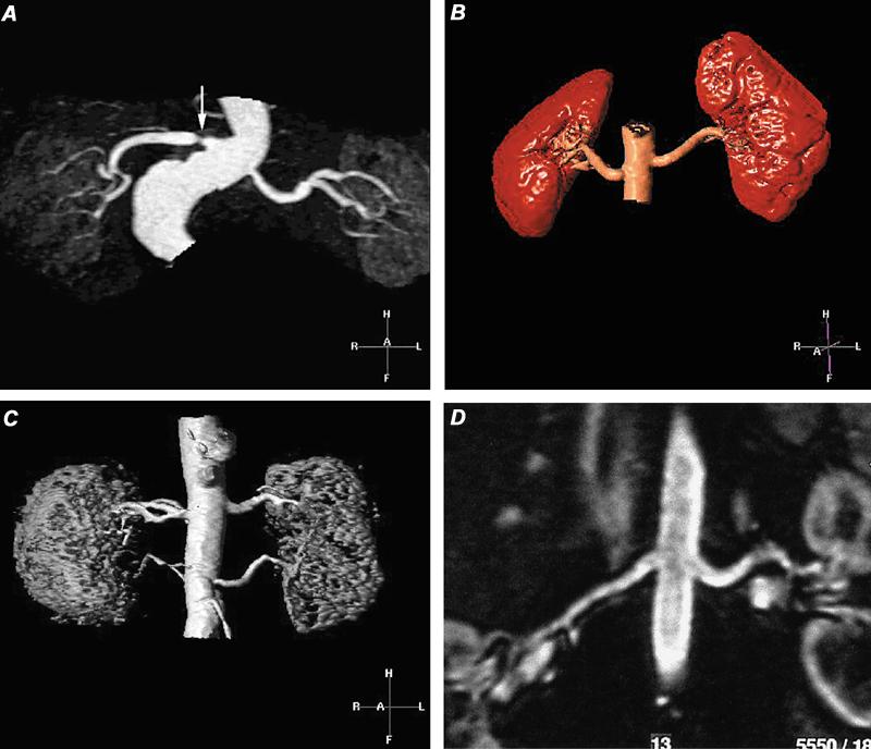
Fig. 18 Contrast-enhanced magnetic resonance angiograms of renal arteries. A) Evaluation by maximum intensity projection (MIP) of hypertensive patient, which discloses a high-grade stenosis (arrow) in the proximal portion of the single right renal artery. The left renal artery appears to be patent. B) Volume-rendered display of normal renal arteries in a renal transplant donor. C) Volume-rendered magnetic resonance angiogram in a hypertensive patient shows patent duplicated renal arteries. D) Curved multiplanar coronal reconstruction with display along the courses of the renal arteries demonstrates patency of the main renal arteries.
