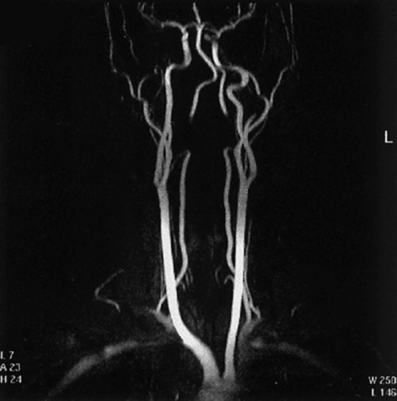
Fig. 19 Contrast-enhanced 3-D display (maximum intensity projection) of the brachiocephalic vessels. This is only 1 of 18 views that are usually displayed and rotated every 10 degrees along the vertical axis of the body, to exhibit all aspects of the cervical and proximal intracranial vasculature. Note: There is signal loss in the distal cervical portions of both vertebral arteries because these segments of the arteries were excluded from the imaging field.
