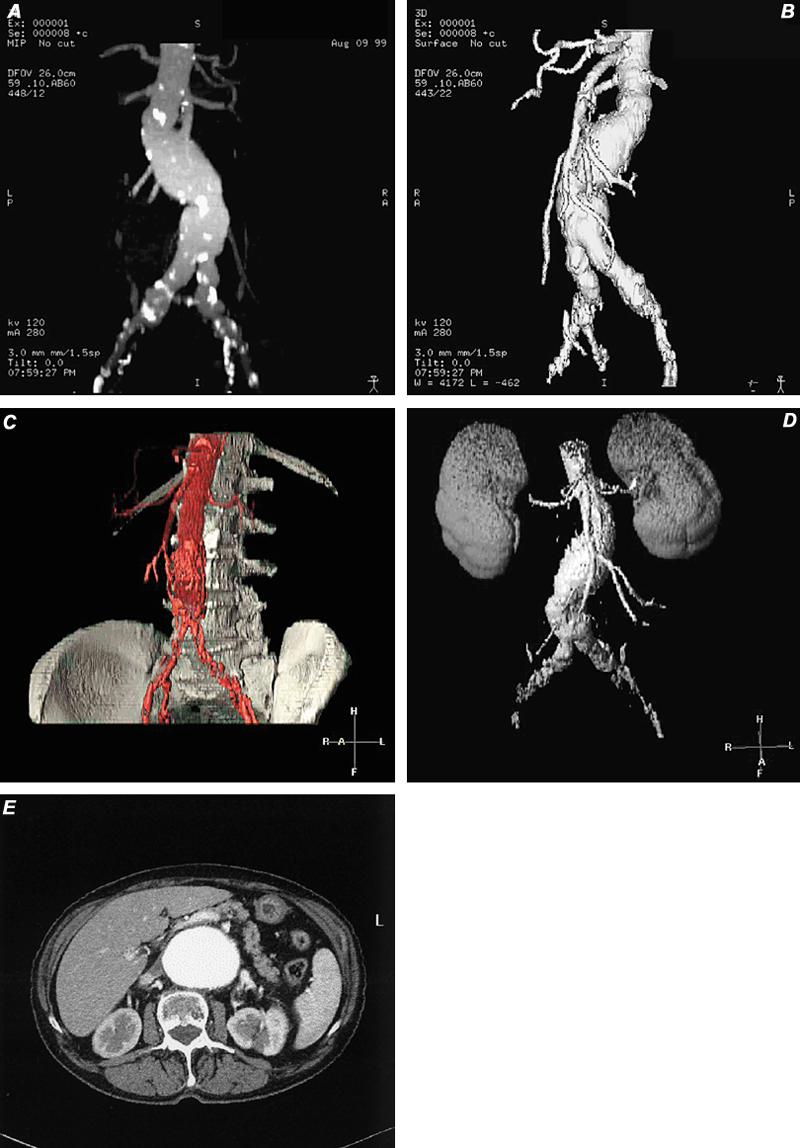
Fig. 20 Types of 3-D display. A) Computed tomographic angiogram using maximum intensity projection (MIP) technique, which discloses a partially calcified fusiform abdominal aortic aneurysm. B) Computed tomographic angiogram with shaded surface display (SSD) depicts the same abdominal aortic aneurysm that appears in image A. Notice that the calcifications are not visible, as they were in MIP imaging. C and D) Two types of volume-rendered images in patients with abdominal aortic aneurysm. E) Axial computed tomographic image of abdominal aortic aneurysm (source image for A-D). Note: A variety of tissues can be presented with this technique.
