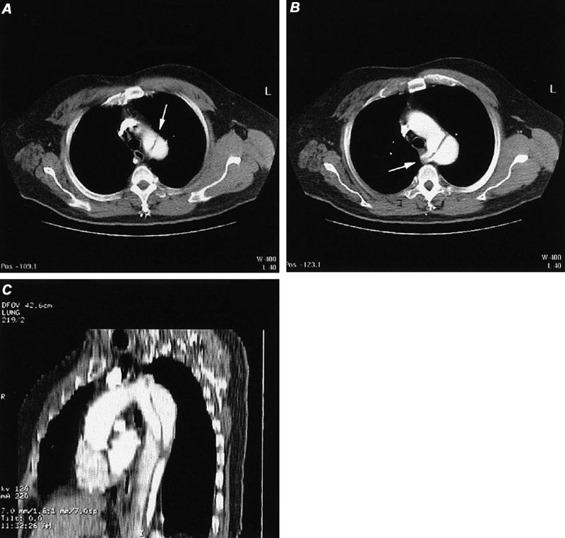
Fig. 22 Computed tomographic angiography of aortic dissection (type B) in a patient with aberrant right subclavian artery. A) Axial image at the apex of the aortic arch discloses an intimal flap in the proximal descending thoracic aorta (arrow). B) Axial image obtained slightly inferior to image A discloses normal appearance of the proximal aortic arch and a dissection flap in the distal aortic arch that extends into the origin of an aberrant right subclavian artery (arrow). C) Reformatted sagittal oblique maximum intensity projection image of thoracic aortic dissection.
