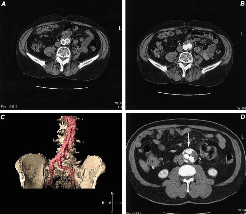
Fig. 23 Computed tomographic angiography in the evaluation of stent-grafts. A) Computed tomographic image of distal abdominal aorta, 6 months following AneuRx™ stent-graft placement. This image was obtained before contrast administration. B) Same location as image A, but during intravascular contrast infusion. Note opacification of both iliac limbs and no contrast extravasation. C) Volume-rendered display of stent-graft discloses no evidence of endoleak. D) Axial computed tomographic image in another patient discloses a leak characterized by contrast pooling (arrow) ventral to the 2 iliac limbs. This was documented to be a non-stent-related endoleak arising from the inferior mesenteric artery. This vessel was embolized successfully.
