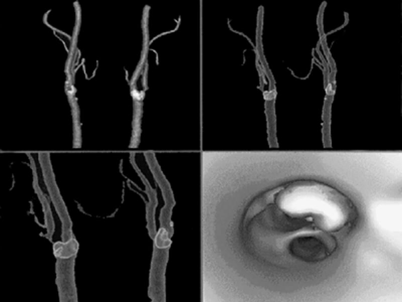
Fig. 28 Computed tomographic angiography of the carotid bifurcations using multi-detector array high-resolution technique. The 1st 3 images are volume-rendered displays of the carotid bifurcations, with calcification in the bifurcations outlined in lighter colors. The last image is a virtual angioscopic display that shows the carotid bifurcation from below, with depiction of a large calcified plaque in the ostium of the internal carotid artery. (Courtesy of General Electric Medical Systems; Milwaukee, Wis)
