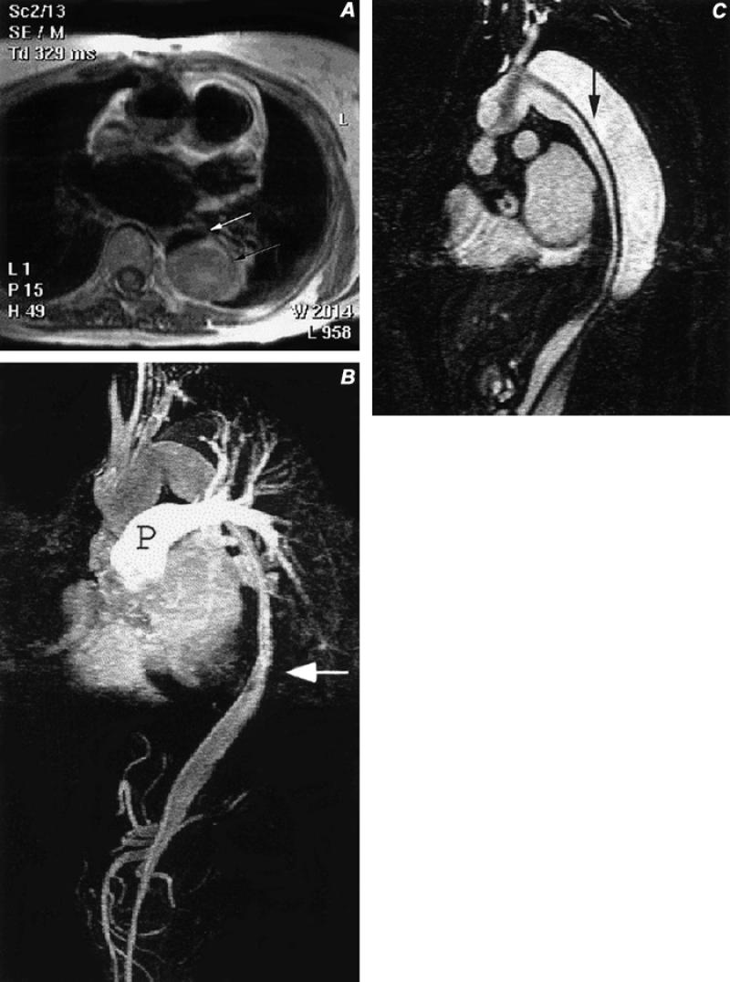
Fig. 5 Type A aortic dissection, after graft repair of the ascending aorta. A) Axial spin-echo image of the mid-thorax with bright signal (black arrow) in the false lumen of the descending thoracic aortic dissection, and with a small, dark, slit-like flow-void in the true lumen (white arrow). B) Sagittal, contrast-enhanced magnetic resonance image of the chest and upper abdomen, disclosing flow in the small ribbon-like true lumen (arrow) with residual contrast in the pulmonary artery (P). Also note opacification of the celiac and superior mesenteric arteries that arise from the true lumen. C) Sagittal image from a contrast-enhanced series of images obtained 15 seconds after image B, showing delayed opacification of the false lumen, which is separated from the true lumen by a dissection flap (arrow) that extends from the proximal aortic arch to below the diaphragm.
