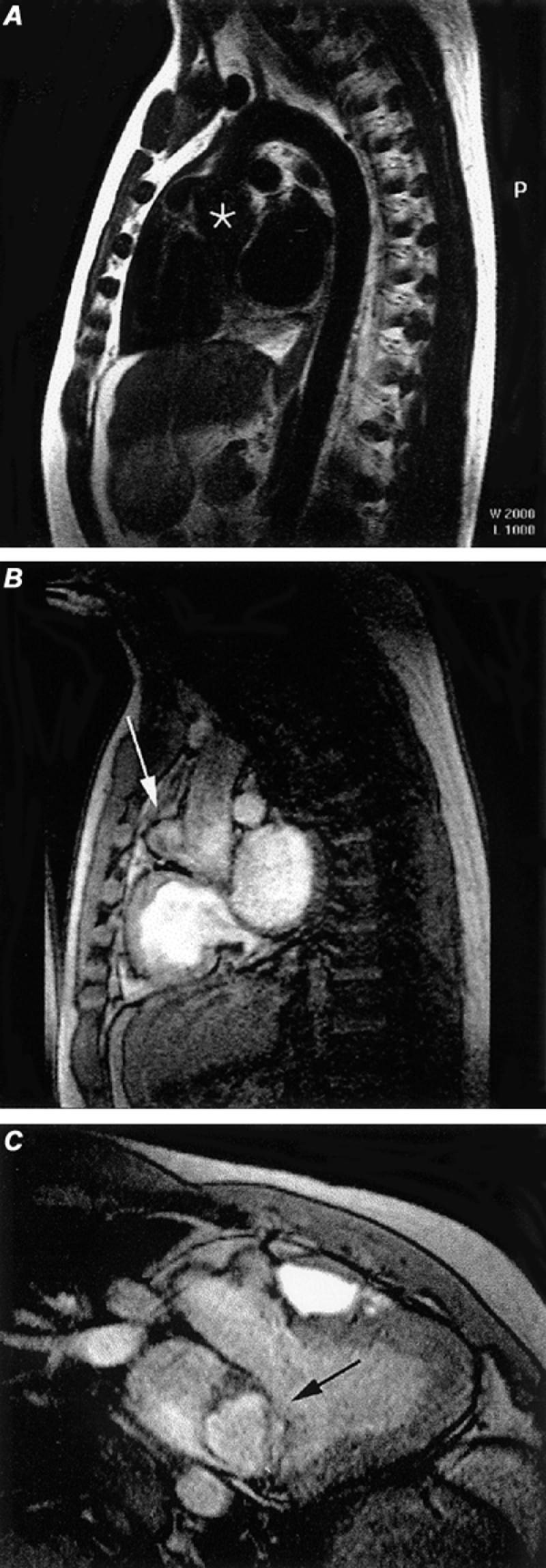
Fig. 6 Sinus of Valsalva aneurysm. A) Oblique spin-echo, black blood image discloses slight dilatation of the aortic root (asterisk). B) Cine gradient-echo sagittal image discloses a small aneurysm (arrow) arising along the anterior aspect of the aortic root. C) Oblique coronal cine gradient-echo image discloses the left ventricular outflow tract and the sinus of Valsalva aneurysm (arrow) arising from the aortic root.
(All images courtesy of Scott D. Flamm, MD)
