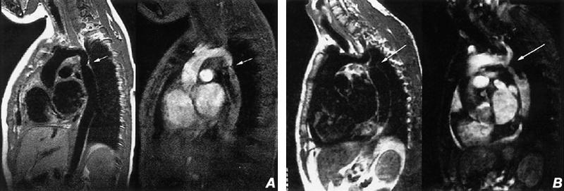
Fig. 8 Coarctation of the aorta. A) Sagittal spin-echo (left) and cine gradient-echo (right) images of a characteristic aortic coarctation with narrowing of the aorta (arrow) just distal to the left subclavian artery. B) Sagittal spin-echo (left) and cine gradient-echo (right) images obtained just slightly to the left of image A, disclosing the high-grade stenosis (arrow) just distal of the origin of the left subclavian artery. (All images courtesy of Scott D. Flamm, MD)
