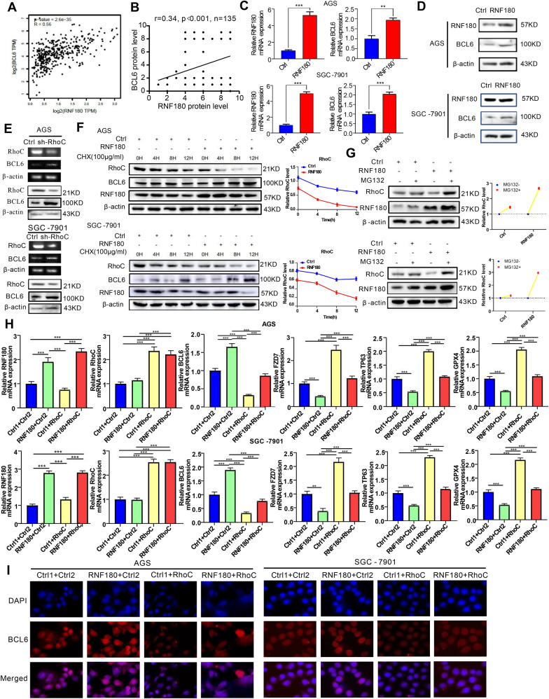
Fig. 7.
Expression of BCL6 in GC cells is mediated by RNF180/RhoC pathway. A Scatter plot shows the positive correlation between RNF180 and BCL6 mRNA expression levels in STAD tumor (N = 408), according to the online GEPIA database; B and the IHC of TMAs (N = 135); C, D Expression levels of BCL6 in AGS and SGC-7901 cells transfected with RNF180 plasmid and control vector were analyzed by qPCR and western blot (**p < 0.01, ***p < 0.001). E Expression levels of BCL6 in AGS and SGC-7901 cells transfected with short hairpin RNA (shRNA) for RhoC or control vector were analyzed by qPCR and western blot. F AGS and SGC-7901 cells were transfected RNF180 plasmid or control vector. After 48 h, cells were treated with 100 mg/ml CHX at the indicated time point. The RhoC protein was measured by western blot. G AGS and SGC-7901 cells were transfected RNF180 plasmid or control vector. After 48 h, cells were incubated with 10 μM MG132 for 24 h.The RhoC protein was measured by western blotting. H RNF180, RhoC, BCL6, FZD7, TP63 and GPX4 mRNA expression in AGS and SGC-7901 cells transfected with control vector (Ctrl1 + Ctrl2, Ctrl1 + RhoC, RNF180 + Ctrl2) or co-transfected RNF180 plasmid and RhoC plasmid (RNF180 + RhoC) (***p < 0.001). I Immunofluorescence staining for BCL6 (red) and 4′,6-diamidino-2-phenylindole (DAPI) (blue) in AGS and SGC-7901 cells transfected with control vector (Ctrl1 + Ctrl2, Ctrl1 + RhoC, RNF180 + Ctrl2) or co-transfected RNF180 plasmid and RhoC plasmid RNF180 + RhoC) (Original magnification, × 400)

