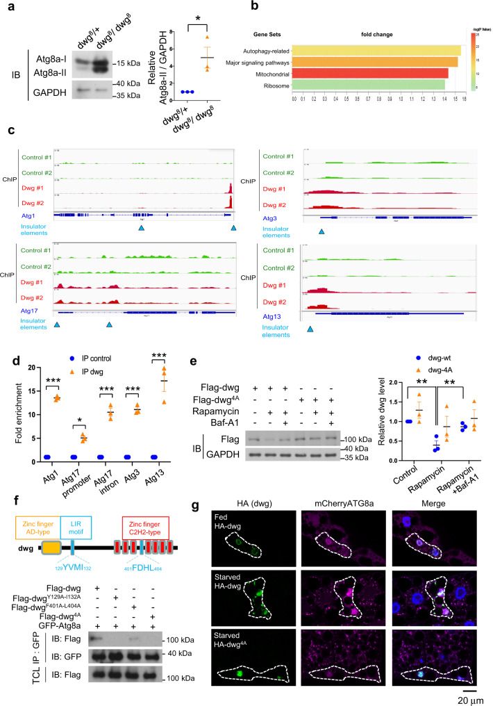
Fig. 4. Dwg is both a negative regulator and a substrate of autophagy.
a Flies homozygous mutant for a loss-of-function allele of dwg exhibit increased autophagy. Control (dwg8/+) and mutant (dwg8/dwg8) larva subjected to immunoblotting with indicated antibodies. Measurements are mean ± SEM of triplicate experiments. Significance determined by two-tailed t-test. *p = 0.031. b Gene group enrichment analysis of ChIPseq data. Bar length, fold change in enrichment. Colors, strength of significance (p-value of -log10 for each term). c, d Dwg binds insulator regions of Atg genes. c Example browser images for Atg1, Atg3, Atg13, and Atg17 from ChIP-seq in S2R + cells expressing Flag-Dwg. Aggregate data from two independent experiments shown. Blue triangles, insulator binding regions. d Dwg occupancy at or near insulator regions of Atg genes revealed by ChIP-qPCR. One-Way ANOVA followed by Tukey’s multiple comparison test to identify significant differences; shown are means ± SEM of three independent experiments; ***P < 0.0001, *P = 0.039. e Autophagic activity regulates Dwg levels. S2R + cells transfected with Flag-dwg or -dwg4A(Y129A-I132A-F401A-L404)) were treated with Rapamycin (autophagy inducer) or Bafilomycin A1 (Baf-A1; lysosomal inhibitor). Dwg and GAPDH levels analyzed by immunoblotting (IB) with indicated antibodies and quantified. One-Way ANOVA followed by Tukey’s multiple comparison test to identify significant differences; shown are means ± SEM of three independent experiments; **P < 0.01; p = 0.0026 (dwg-wt control v.s. Rapamycin). p = 0.0089 (dwg-wt Rapamycin v.s. Baf-A1). f Mapping Dwg-Atg8a interaction sites. Schematic of domain structures and LIR (LC3-interacting region) motifs of Dwg. S2R + cells transfected with GFP-Atg8a, Flag-tagged dwg, or Flag-tagged dwg with mutations in LIR motif (dwgY129A-I132A, dwgF401A-L404A, or dwg4A(Y129A-I132A-F401A-L404)) for 48 h followed by immunoprecipitation with anti-GFP nanobody. Immunoprecipitated proteins and total cell lysates analyzed by immunoblotting with indicated antibodies. g Disruption of Dwg-Atg8a interaction inhibits autophagy. Clonally expressed HA-tagged Dwg is nuclear under fed conditions (green); upon starvation, it is detected in cytoplasm and co-localizes with autophagosomes labeled by mCherry-Atg8a (magenta). A version of Dwg with mutations in Atg8a-binding sites (dwg4A) remains nuclear and inhibits autophagy. Fat body cells stained with DAPI (blue). Experiments repeated three times independently with similar results. Scale bar: 20 μm. Source data are provided as a Source Data file.

