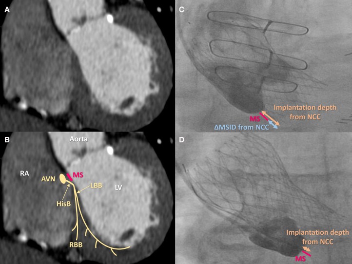
Figure 1.
Schematic illustration of cardiac conduction system on cardiac computed tomography and aortic angiogram. Reconstructed coronal view of cardiac computed tomography (A), schematic illustration of cardiac conduction system on cardiac computed tomography (B), aortic aortogram in patient with deep valve implantation (C), and aortic aortogram in patient with shallow valve implantation (D). AVN, atrioventricular node; HisB, His bundle; LBB, left bundle branch; LV, left ventricle; MS, membranous septum; NCC, non-coronary cusp; RA, right atrium; RBB, right bundle branch; ΔMSID, the difference between the MS length and the implantation depth.

