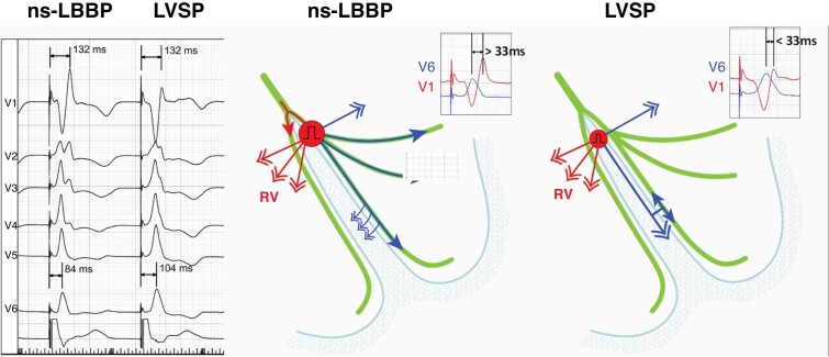
Figure 25.
Illustration of effect of ns-LBBP and loss of conduction tissue capture resulting in LVSP with myocardial capture only, on V6–V1 inter-peak interval, occurring during a threshold test. R-wave peak time in V1 reflects right ventricular activation that depends on transseptal conduction and is not affected by conduction tissue capture. RWPT in V6 reflects activation of the lateral wall of the left ventricle and is not influenced by loss of septal myocardial capture. Consequently, a longer V6–V1 interval is observed with left conduction system capture. Modified, with permission, from Jastrzebski et al.20

