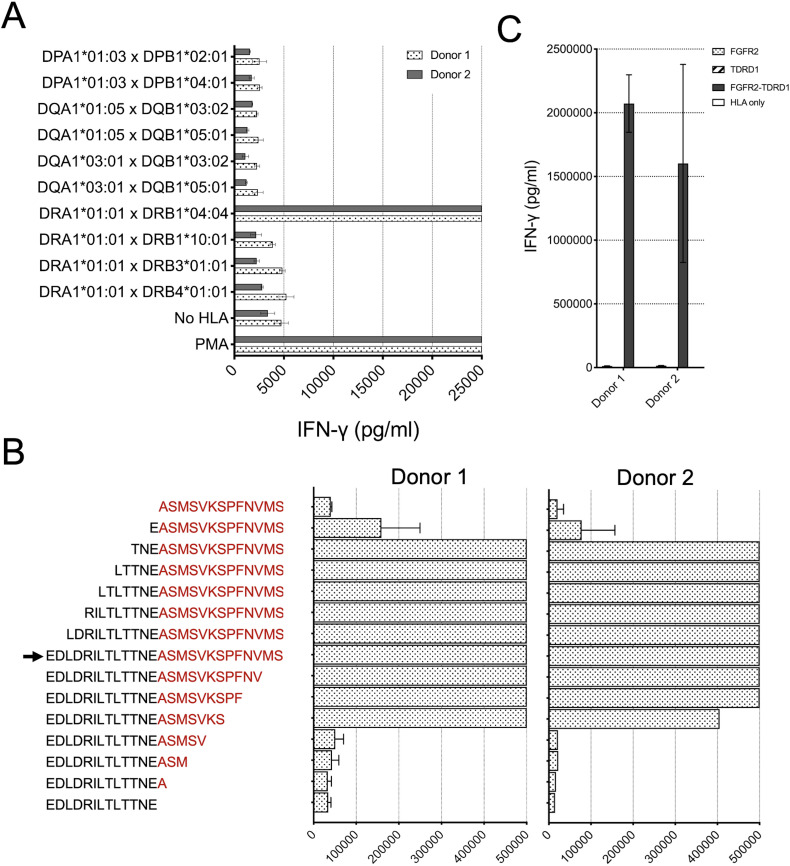
Figure 4.
TCR1 demonstrated HLA-DRB1*04:04-restricted recognition of FGFR2-TDRD1 peptide, which could be derived from the breakpoint region in the FGFR2-TDRD1 minigene. (A) TCR1-transduced T cells from two healthy donors were co-cultured with COS-7 cells, which were first transfected with pairs of plasmids encoding all MHC class II molecules detected in patient 1’s tumors, and then pulsed with the FGFR2-TDRD1 peptide. The following day, IFN-γ production was measured using an IFN-γ electrochemiluminescence assay. Mock-transfected COS-7 cells (No HLA) were used as a negative control; PMA/ionomycin (PMA) was used as a positive control. (B) Same TCR1-transduced T cells were co-cultured with patient 1’s B cells that were pulsed with peptides derived from the FGFR2-TDRD1 26-mer (arrowhead) by serial truncations of the FGFR2 (black) or TDRD1 portion of the fusion (red). IFN-γ production was measured the following day. (C) TCR1-transduced T cells were co-cultured with COS-7 cells that were transfected with plasmids encoding HLA-DRA1*01:01 and DRB1*04:04 molecules and a plasmid encoding either the breakpoint region of the FGFR2-TDRD1 protein, or the corresponding portions of normal FGFR2 or TDRD1 proteins. IFN-γ production was measured via electrochemiluminescence. COS-7 cells transfected only with the HLA plasmids were used as a negative control. For all experiments in this figure, bar graphs represent average reads from duplicate co-culture wells; error bars represent SD. FGFR2, fibroblast growth factor receptor 2; HLA, human leukocyte antigen; IFN-γ, interferon gamma; MHC, major histocompatibility complex; PMA, phorbol 12-myristate 13-acetate; TCR, T-cell receptor; TDRD1, tudor domain-containing 1.

