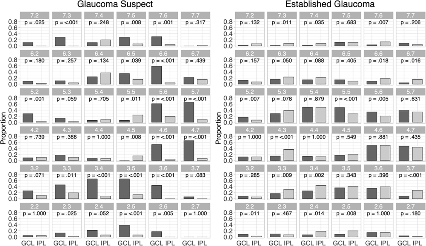
Figure 3.

The proportion of eyes with significantly negative rates of change for ganglion cell layer (GCL) and inner plexiform layer (IPL) for each superpixel for the glaucoma suspect (left) and established glaucoma (right) cohorts (dark gray bars: GCL; light gray bars: IPL). P values for comparison between proportions are based on McNemar’s test.
