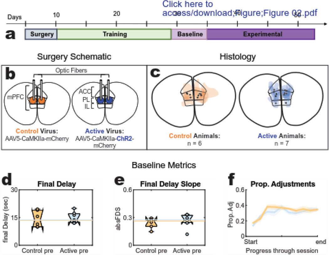
Figure 2: Task stages, Histology, and baseline behaviour.
(a) Task Stages: Following surgery recovery, rats entered step-wise training blocks that introduced them to the DD task until they were running reliably with a head tether. Baseline behavior was collected for 6 days prior to the 18 day opto-delivery experimental sequence (b) Surgery schematic: the prelimbic (PL) region was targeted for bilateral injection of either an active (opsin expressing) or control (opsin absent) virus and optic fiber implantation. (c) Histology: target variation for viral expression (color splotches) and fiber tips (black marks) of each subject in either cohort (three rats presented with unilateral expression and will be identified in the results with dashed lines). (d-f) Baseline session-level metrics: “pre” data was collected during the baseline stage that confirmed both cohorts were preforming sufficiently on the task by achieving comparable (d) final delay values, (e) final delay slopes, and (f) proportion of adjustment laps.

