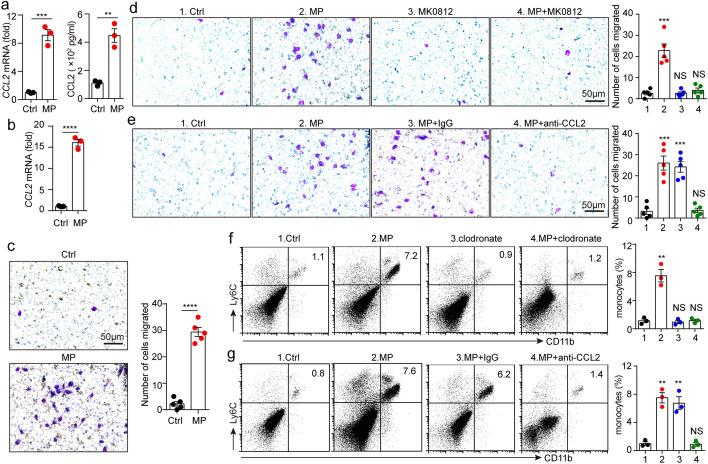
Fig. 3.
T-MPs induce CCL2 upregulation in macrophages. a BALB/c mouse peritoneal macrophages were cultured with H22-MPs. After 24 or 72 h, RNAs or cultured medium was collected respectively. Then, the expression of CCL2 was measured using real-time PCR or ELISA. b BALB/c mice were intramuscularly inoculated with H22-MPs or PBS. 24 h later, CCL2 expression of isolated monocytes from thigh muscles was measured by real-time PCR. c BALB/c mouse bone marrow-derived macrophages (BMDMs) were co-incubated with H22-MPs in a 24-well plate, and isolated monocytes from bone marrow cells were cultured in the upper chamber, followed by monocyte migration assay after 48 h. d BALB/c mouse BMDMs were co-incubated with H22-MPs, and monocytes were cultured with or without CCR2 antagonist MK0812 in the upper chamber, followed by monocyte migration assay after 48 h. e BALB/c mouse BMDMs were treated with H22-MPs and CCL2 neutralizing antibody, and monocytes were cultured in the upper chamber, followed by monocyte migration assay after 48 h. f BALB/c mice were subjected to clodronate liposomes by tail vein twice a week for 3 weeks, then mice were treated with H22-MPs via i.m. injection. After 48 h, the proportion of monocytes in muscles around the femur was measured by flow cytometry. g BALB/c mice were given i.m. injection with H22-MPs, PBS control or intraperitoneally pretreatment with anti-CCL2 neutralizing antibody. After 48 h, the proportion of monocytes in muscles around the femur was measured via flow cytometry. All of experimental groups compared with control group. Mean ± s.e.m. is represented in the data and two-tailed unpaired Student's t test was used to statistically analyze the P values. **P < 0.01; ***P < 0.001; ****P < 0.0001; NS, not significant

