FIGURE 4.

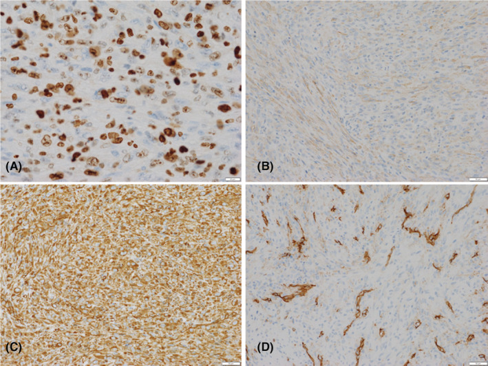
Immunohistochemical staining. Neoplastic cells strongly stained for MIB‐1 (A), α‐smooth muscle actin (B), vimentin (C), and CD 34 (D), and are focally positive.

Immunohistochemical staining. Neoplastic cells strongly stained for MIB‐1 (A), α‐smooth muscle actin (B), vimentin (C), and CD 34 (D), and are focally positive.