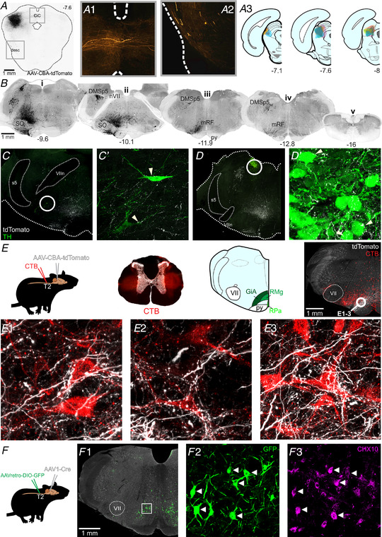
Figure 5. Anterograde labelling from the cl‐dSC.

A, example of AAV‐CBA‐tdTomato injection site in the cl‐dSC; fibres emerging from the injection site projected to the contralateral colliculi via the commissure of the inferior colliculus (CiC, A1) or followed a descending trajectory via the lateral midbrain (desc, A2). A3, composite diagram showing distribution of injection sites in six experiments. B, histological sections from one experiment, illustrating the brainstem distribution of tdTomato labelling. The descending bundle split into two tracts in the pons, a more substantial ventral bundle that surrounded the superior olive on all sides (SO, Bi), and a lesser and exclusively ipsilateral dorsal tract that innervated the dorsomedial spinal trigeminal nucleus (DMSp5, Bii) with large‐calibre fibres. The ventral branch occupied a column extending from the ventral surface to an apex at the locus coeruleus, was sparsely mirrored on the contralateral brainstem, and comprised both large‐ and fine‐calibre fibres and putative terminals. Caudal to the facial nerve (nVII, Bii) the ventral branch was most densely concentrated in the medullary reticular formation (mRF, Biii) dorsal to the pyramidal tract (py) at the level of the caudal pole of the facial nucleus. Labelled fibres were infrequently encountered in the caudal medulla (Biv) and virtually absent from spinal sections (Bv). Coordinates indicate the rostrocaudal position with respect to the bregma. Putative targets of the cl‐dSC outputs included rare tyrosine hydroxylase (TH) immunoreactive neurons in the A5 (C) and A6 (D) cell groups, and a conspicuous terminal field in the gigantocellular alpha region of the reticular formation (GiA, E). Putative cl‐dSC projections to spinally projecting neurons in the GiA were identified in experiments in which anterograde cl‐dSC labelling was combined with retrograde labelling of spinally projecting neurons by microinjection of cholera toxin B (CTB) at the thoracic intermediolateral cell column: top row of panel E shows a schematic of the experimental strategy; brightfield transverse thoracic spinal cord section superimposed with CTB injection sites in red; and an atlas plate highlighting the GiA, midline raphe magnus (RMg) and pallidus regions (RPa) with a low‐power image showing the distribution of CTB‐labelled cell bodies (red) and cl‐dSC terminals (white). E1–3, high‐power confocal images from the circled region show close apposition between cl‐dSC terminals and spinally projecting GiA neurons. F, similar results were obtained using AAV1‐mediated trans‐synaptic tagging: spinally projecting neurons were transduced by injection of Cre‐dependent AAVretro‐hSyn‐DIO‐GFP in the thoracic spinal cord, GFP expression controlled by the trans‐synaptic trafficking of AAV1‐hSyn‐Cre from cl‐dSC. F1, GFP‐immunoreactive neurons were exclusively found in GiA and included CHX10‐immunoreactive V2A neurons (arrowheads) and CHX‐10 negative cells (F2 and 3). [Colour figure can be viewed at wileyonlinelibrary.com]
