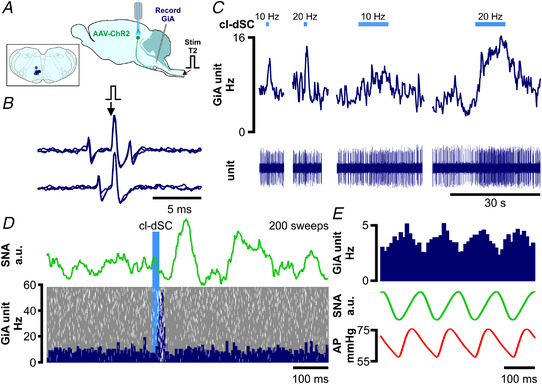
Figure 8. Optogenetic cl‐dSC stimulation activates spinally projecting cardiovascular and non‐ cardiovascular GiA neurons.

A, extracellular recordings were made from bulbospinal GiA neurons under urethane anaesthesia after transduction of cl‐dSC neurons with ChR2 and fibre optic implantation; inset shows recording sites of seven neurons. B, spinally projecting neurons exhibited constant‐latency antidromic spikes in response to electrical stimulation of the T2 spinal cord (top trace) which collided with spontaneous orthodromic spikes (lower trace). C, cl‐dSC stimulation evoked frequency‐dependent increases in baseline activity: trace shows typical responses to short (1 s) and long (10 s) trains of stimulation at 10 and 20 Hz (same neuron as B). D, low‐frequency cl‐dSC stimulation evoked short‐latency response in the same neuron (laser‐triggered peristimulus time histogram (dark blue) with overlaid raster (white spikes, grey background, lower panel) that preceded simultaneously recorded splanchnic sympathetic responses (green trace, upper panel)). E, putative sympathetic premotor neurons were identified by covariation of spontaneous neuronal activity (top trace) with SNA (green) and pulsatile arterial pressure (lower trace). [Colour figure can be viewed at wileyonlinelibrary.com]
