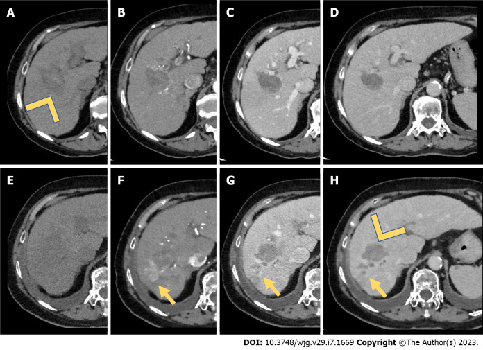
Figure 1.
Computed tomography study for assessment of treatment response (after microwave ablation). A: A 65-year-old male underwent microwave ablation of a hepatocellular carcinoma located in the V-VIII hepatic segment. Computed tomography scans were acquired after 2 wk of treatment. A large hypoattenuating area in the unenhanced (arrowhead) phase located in the V-VIII hepatic segment represented the treatment zone; B-D: During the dynamic study, no enhancement during the arterial phase (B) was seen, underlying the complete treatment response. Also, during the portal venous phase (C) and delayed phase (D) no wash-out was seen; E-H: After 1 year, the area of treatment was less hypoattenuating in the unenhanced phase (E), with a pseudonodular peripheral area of hypervascularization during the arterial phase (F, yellow arrow), with a wash-out during the portal venous and delayed phases (G and H, yellow arrow). On the other hand, the area of treatment did not show any arterial phase hyperenhancement or wash-out (H, arrowhead). The final diagnosis was hepatocellular carcinoma recurrence after microwave ablation (yellow arrows).

