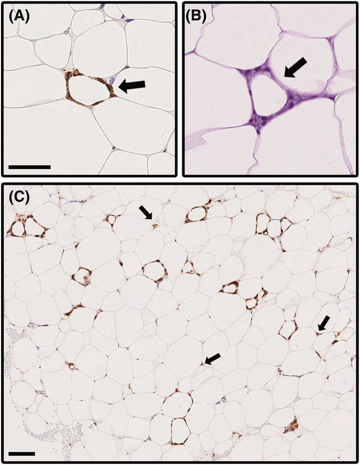
FIGURE 1.

Microphotographs illustrating crown‐like structures (CLS) and dispersed CD68‐positive cells in subcutaneous adipose tissue of participants with overweight. Identification of CLSs is facilitated with (A) CD68 staining (arrow) as compared with (B) H&E staining (arrow). Scale bar = 50 μm. (C) Sometimes CLSs were present at a high density as shown by CD68 staining. A 40‐year‐old woman with BMI 44.9 kg/m2 and no comorbidities had CLS density of 14.33 per 1000 adipocytes, the highest count found among the subjects. A particularly high‐density region is shown in the image. This patient's CLS density had decreased to 1.23 per 1000 adipocytes and BMI decreased to 37.7 kg/m2 at 12 months after the bariatric surgery. Arrows point to some CD68‐positive cells dispersed outside CLSs. Scale bar = 100 μm [Color figure can be viewed at wileyonlinelibrary.com]
