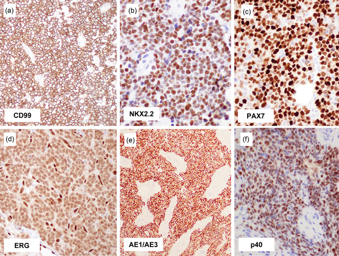
Figure 2.

Immunohistochemical findings of Ewing sarcoma. Ewing sarcoma virtually always expresses CD99, often in a diffuse strong, and membranous pattern (a). Most examples are positive for NKX2.2 (b) and PAX7 (c), irrespective of histological pattern and fusion gene variant, and this combination of staining is highly specific for Ewing sarcoma. Diffuse ERG expression indicates the presence of EWSR1::ERG fusion (d). Keratin can be expressed in Ewing sarcoma, which can be rarely diffuse and strong (e), mimicking epithelial tumors. Adamantinoma‐like Ewing sarcoma expresses high‐molecular‐weight cytokeratin and p40 (f), consistent with squamous differentiation.
