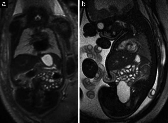
Figure 9.

(a) Coronal T2‐weighted magnetic resonance image in a 32 + 2‐week fetus, displaying fluid‐filled stomach and bowel loops. (b) Sagittal steady‐state free‐precession (SSFP) image in a 35 + 6‐week fetus, showing in addition the fluid‐filled urinary bladder. Note the hyperintensity of the heart in this image in contrast to the T2‐weighted image (a).
