Figure 3.

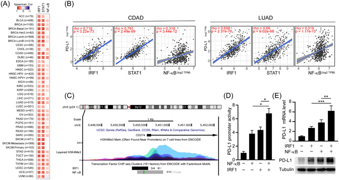
NF‐κB/IRF1 axis upregulates PD‐L1 expression. (A) Spearman correlation analysis of PD‐L1 with IRF1, STAT1, and RELA (NF‐κB) in the TCGA pan‐cancers. (B) Spearman correlation analysis of PD‐L1 with IRF1 and STAT1 in the TCGA colon cancer (COAD; n = 460) and lung cancer data set (LUAD; n = 585). (C) ENCODE data displayed ChIP‐sequencing signals for the occupancy of IRF1, RELA, and histone H3K4me3 around the transcription start sites of PD‐L1. (D) Promoter‐luciferase reporter activity of PD‐L1 (−373/+328) by overexpression of NF‐κB and IRF1. Statistical method: one‐way ANOVA, Tukey post hoc tests, *p < 0.05. (E) Effects of IRF1 and NF‐κB on the mRNA expression (upper panel) and protein expression (lower panel) of PD‐L1 in A549 cells were analyzed by RT‐qPCR and western blotting, respectively. Statistical method: one‐way ANOVA, Tukey post hoc tests, **p < 0.01, ***p < 0.001. ANOVA, analysis of variance; ChIP, chromatin immunoprecipitation; IRF1, interferon regulatory factor 1; LUAD, lung adenocarcinoma; mRNA, messenger RNA; NF‐κB, nuclear factor‐κB; PD‐L1, programmed death ligand‐1; RT‐qPCR, real‐time quantitative polymerase chain reaction; TCGA, The Cancer Genome Atlas.
