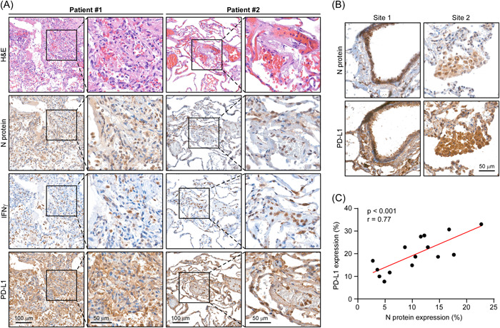
Figure 6.

PD‐L1 correlates with N protein in human COVID‐19 lung tissues. (A) Representative microscopic images of N protein, IFN‐γ, and PD‐L1 expression in human COVID‐19 lung tissues. (B) Representative microscopic images of N protein and PD‐L1 expression in human COVID‐19 lung tissues. (C) Spearman's rank correlation analysis between the protein expression levels of N protein and PD‐L1 in (B). (3 fields per lung tissue; total 15 fields). IFN‐γ, interferon‐γ; PD‐L1, programmed death ligand‐1.
