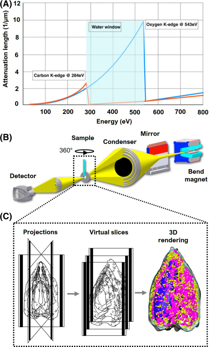
FIGURE 1.

Overview of imaging cells with SXT. (A) “Water window” (light blue): region of the spectrum where the X‐ray absorption is less attenuated by water, 23 giving rise to natural contrast of cell structures in SXT. (B) Schematic representation of XM‐2, the soft X‐ray microscope located at Advanced Light Source, Lawrence Berkeley National Laboratory. Details about the microscope can be found on the National Center for X‐ray Tomography website (https://ncxt.org/) and in previous articles 37 ; (C) Specimen acquisition: (Left) single projections at incremental degrees of rotation are collected, aligned around the rotation axis, and reconstructed; (Center) virtual sections, or orthoslices, of the reconstructed tomograms; (Right) 3D volume of the reconstructed cell with select organelles segmented and color‐coded (reconstructed data adapted from White et al., 2020 38 ).
