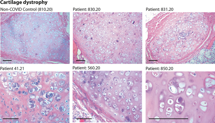
Figure 2.

Histology of bronchial cartilage. Haematoxylin and eosin (H&E) staining is shown for five previous COVID‐19 patients and one normal control who died from non‐COVID pneumonia. In previous COVID‐19 patients, chondrocytes appear randomly clustered. Several cells show marked basophilic degeneration, with increased size, dysmorphic nuclear structure, and peripheral halos. In several instances, the chondrocytes have become anucleated. Scale bars: 100 μm.
