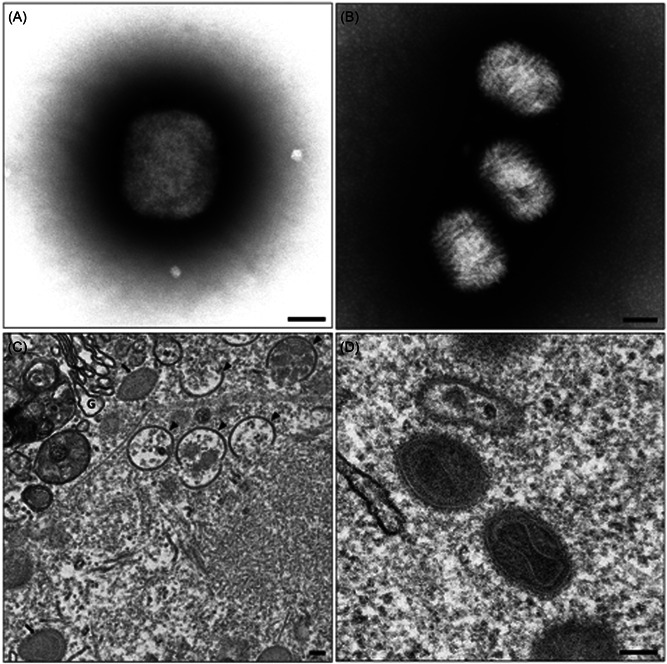
Figure 3.

Transmission electron micrographs provided by Dr. Jason Roberts. Negative‐stained Monkeypox virus (MPXV) positive cell culture supernatant, showing an approximately 250 nm × 300 nm brick‐like particle indicative of the genus Orthopoxvirus, (B) Negative‐stained molluscum contagiosum positive material showing multiple 190 nm × 260 nm ovoid particles with a woven appearance of surface tubules characteristic of the genus Molluscipoxvirus, (C) cytoplasmic region of an MPXV infected Vero cell showing a typical virus factory or virosome, intracellular mature virus particles (black arrows) and intermediate crescent stage immature virus particles (black arrowheads) are visible, G = Golgi body, (D) multiple intracellular mature MPXV particles can be seen with clearly defined dumbbell‐shaped core and striated palisade layer between inner and outer membranes. Scale bar = 100 nm.
