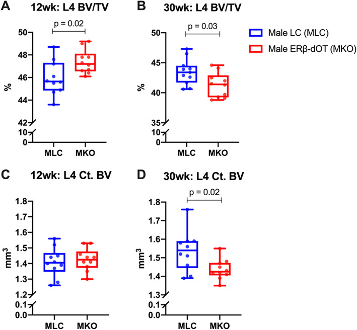
Fig. 1.

Bone morphology analysis of L4 in 12‐week‐old (young) and 30‐week‐old (adult) male littermate controls (LC) and ERβ‐dOT (knockout [KO]) mice. The lumbar vertebral body (L4) of male LC (MLC, blue) and ERβ‐dOT (MKO, red) mice at 12 weeks (A, C) and 30 weeks of age (B, D) were analyzed by micro‐CT. L4 trabecular bone volume fraction (BV/TV; A, B) and cortical bone volume (Ct.BV; C, D) of 12‐week‐old and 30‐week‐old MLC and MKO are presented as box plots with median and interquartile ranges (IQR; 25th to 75th percentile) including all data points (n = 10–12 per group). Significant differences in L4 bone morphology between genotypes were tested by Student's t test (p < 0.05). Specific p values are shown when p < 0.05.
