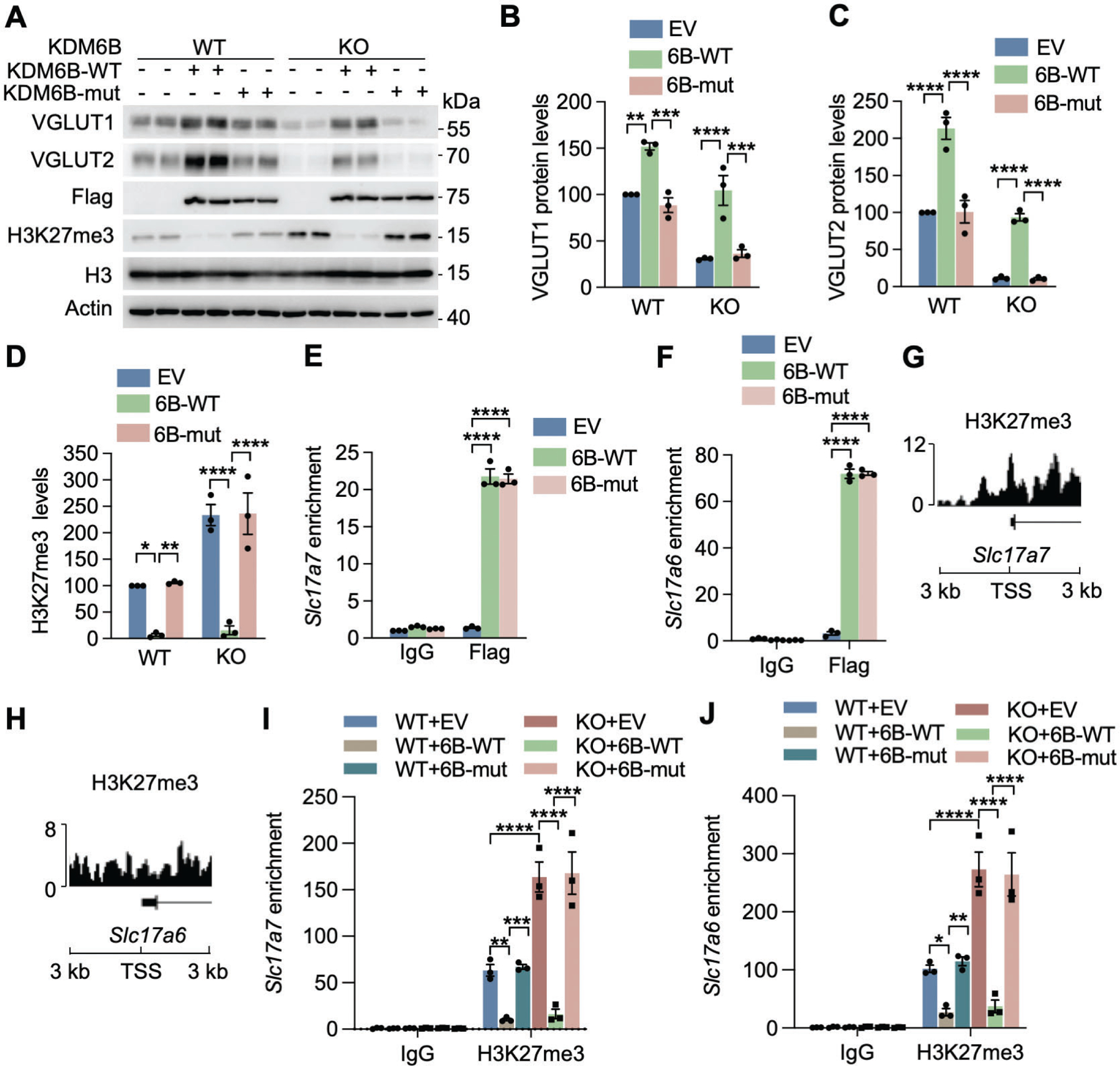
Fig. 4. KDM6B induces VGLUT1 and VGLUT2 expression through its H3K27me3 demethylase activity.

(A-D) Representative immunoblot (A) of VGLUT1 and VGLUT2 expression in WT and KDM6B KO neurons (DIV 14) expressing empty vector (EV) or KDM6B-C-WT or catalytically mutant (mut). The intensity of VGLUT1 (B), VGLUT2 (C), and H3K27me3 (D) bands was quantified and normalized to β-actin. n = 3 biological replicates in each group. One-way ANOVA with Tukey’s multiple comparison test. (E, F) ChIP-qPCR analysis of WT and mut KDM6B enrichment at the promoters of Slc17a7 (E) and Slc17a6 (F) in neurons (DIV 14). n = 3 biological replicates in each group. Two-way ANOVA with Sidak’s multiple comparison test. (G, H) H3K27me3 ChIP-seq peaks at the transcriptional start sites (TSS; ±3 kb) of Slc17a7 (G) and Slc17a6 (H) in neurons. Data were retrieved from GSM2800528. (I, J) ChIP-qPCR analysis of H3K27me3 at the promoters of Slc17a7 (I) and Slc17a6 (J) in WT and KDM6B KO neurons (DIV14) expressing EV or KDM6B-C-WT or mut. n = 3 biological replicates in each group. Two-way ANOVA with Tukey’s multiple comparison test. Data were presented as mean ± s.e.m. *p < 0.05; **p < 0.01; ***p < 0.001; ****p < 0.0001.
